Products /
CytoCHECK SPAchip® Calcium and pH Multi-Detection Kit
CytoCHECK SPAchip® Calcium and pH Multi-
CytoCHECK SPAchip® Calcium and pH Multi-Detection Kit allows continuous, simultaneous, and precise monitoring and measuring of both cytosolic Calcium and intracellular and extracellular pH levels by changes in fluorescence intensity. This facilitates a more comprehensive study of living single-cell physiology. The main advantage of this product is that combines the two pH and calcium detection technologies in one single SPAchip, enabling real-time, simultaneous, and accurate monitoring of intracellular and extracellular pH and calcium levels in single living cells. These cell-based assays are useful for studying and tracking important biological processes as well as for evaluating anti-cancer drug treatments.
Kit box contains:
- 1 ASSAY SPAchip® eppendorf tube (Quantity: 2.5 x10^6 ASSAY SPAchip®)
- 1 Control SPAchip® eppendorf tube (Quantity: 2.5 x10^5 CONTROL SPAchip®)
- 1 ASSAY buffer tube (Quantity: 5mL)
- Quick start guide
- CytoCHECK SPAchip® Multi-Detection kits are optimized for its use with confocal microscopes and HCS/HCA analyzers with 20X or over magnification objectives, yet widefield fluorescence microscopes and imaging system with fixed wavelength filters can be also used. Flow cytometers have additionally been validated to analyze intra and extracellular SPAchips.
- SPAchip® kits can be handled as common fluorophores and chemical probes for cell biology. After resuspending them in the assay buffer (a SPAchip®-to-cell ratio of 2:1 is recommended), an overnight incubation is required to allow SPAchip® to be incorporated in the cytosol. SPAchip® will remain in the cytosol for longer than one month to monitor cell culture progression.
What are the key features?
- SPAchip® assay kits are novel cell-based assays for living single-cell that bring together the fields of nanotechnology and cell biology.
- CytoCHECK SPAchip® Calcium and pH Multi-Detection Kit enables continuous, simultaneous, and accurate monitoring of intracellular and extracellular pH and Calcium levels in living cells, enabling a more comprehensive study of cell health and physiology.
- CytoCHECK SPAchip® Calcium and pH Multi-Detection kits are composed of fluorescently labeled silicon microparticles -SPAchips®- that can be internalized in cultured cells to monitor changes in specific intracellular analyte concentrations for long periods of time.
- 2-sensors-in-one SPAchip: the main advantage of this product is the combination of our two pH and calcium detection technologies in a single SPAchip device, simplifying cell-based assays by using one single technique. Experimental readouts obtained from the same single cell diminish variability and allow to establish more reliable correlations.
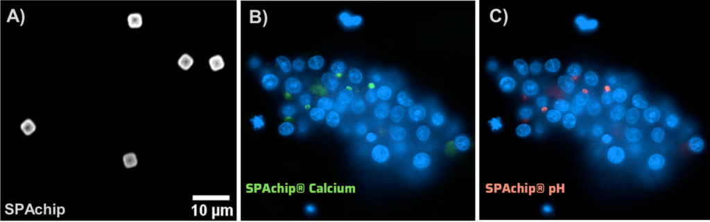
Figure 1: A) Fluorescence signal of SPAchips in an imaging plate with cell culture medium. B) Representative image of SH-SY5Y cells with internalized multiplex SPAchips excitating at 488 nm and emitting at 520 nm. C) Representative image of SH-SY5Y cells with internalized multiplex SPAchips excitating at 546 nm and collecting emission at 610 and 707 nm.
Technical specifications
Product code | M-001-PC | |
Amount | ~2.5 millions of SPAchips | |
Applications | Cell viability, proliferation, cell image acquisition | |
Assay time | 30 minutes | |
Assay type | Living single-cell based | |
Solubility | Soluble in assay buffer (aqueous) | |
Analyte | Calcium | pH |
Detection method | Green | Red fluorescence / Ratiometric curve* |
Fluorescence | λex: 488 nm; λem: 520 nm | λex: 546 nm; λem: 610 and 707 nm |
Measuring range | 10 -1000 μM | 4.5 – 9.0 |
Compatible Platforms | Fluorescence microscopy, HCS/HCA platforms (20x magnification and over) and flow cytometry | |
Sample type | Adherent cells, suspension cells |
Image Acquisition and Quantitative Analysis
CytoCHECK SPAchip® kits are designed to dynamically shift fluorescence intensity values in response to intracellular changes in analyte concentration. Fluorescence intensity to quantify calcium levels is measured by exciting the live cell sample at 488 nm and detecting fluorescence emission at 520 nm. To quantify intracellular and extracellular pH, fluorescence intensity is measured by exciting at 546 nm and emitting at 610 nm and 707 nm. Relative intensity units depend on the instrumental setup (excitation power, detector efficiency, etc.). To obtain quantitative data, a calibration curve needs to be analyzed in the same run. Data acquisition can be adapted to fluorescence microscopy and flow cytometry.
Additional detection channels, such as transmitted light, are suggested to better identify and localize the cells and SPAchips of interest.
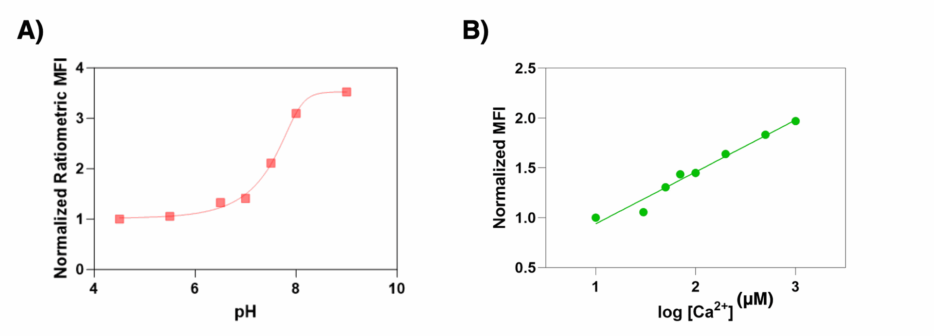
Figure 2: CytoCHECK SPAchip® Calcium and pH Multi-detection Kit at different calcium ionophores and chelators and at different pH conditions using commercial calibrators A) Graph showing ratiometric normalized fluorescence intensity values at different pH. Ratiometric values were obtained by dividing λemi2/ λemi1 emission signals in HCS-Operetta equipment with the excitation in the range λexc=545/15 nm. B) Extracellular calcium calibration curve.
SPAchip® fluorescence signal quantification can be adapted to any analysis image software. Object segmentation should be performed to identify SPAchips inside cellular region to every sample. Fluorescence intensity units can be exported as a spreadsheet to plot graphical summaries of the results. To quantify intracellular pH, use the fluorescence of the calibration wells to plot ratiometric fluorescence units vs calibration pH. Interpolate the values of the sample wells to obtain the intracellular pH value of the sample wells.
* Contact A4cell staff for assistance with imaging setup and data analysis. Ask for our cell biology image analysis guide at info@ols-bio.de


What would you use SPAchip® technology for?
Tell us and help understanding and developing our product