Spheroids
Spheroids
Th CERO 3D's advanced capabilities empower scientists to analyze complex cellular mechanisms, such as the role of lysophosphatidic acid in ovarian cancer cell survival and proliferationCERO 3D enables researchers to make groundbreaking discoveries that could lead to new therapeutic strategies for cancer treatment. advent of long-term three dimensional cell culture holds a great promise in disease modeling and drug discovery. The cells kept in a 3D environment have the ability to mimic tissue-like structures more efficiently than in traditional 2D monolayer cultures. However, many scientist are struggling with many technical limitations when working with spheroids in long-term cultures.
The CERO 3D Incubator & Bioreactor is a revolutionary technology enabling scientists to perform experiments they were not able to do before.
- No necrosis and apoptosis
- Viability in long-term culture > 80 days
- Improved maturation
- Long-term proliferation
- High homogeneity
- High yield
Spheroids from HepG2 cells (hepatocyte cell line) cultivated in CERO 3D for >80 days.


New Insights into Cancer Reserach
CERO 3D's advanced capabilities empower scientists to analyze complex cellular mechanisms, such as the role of lysophosphatidic acid in ovarian cancer cell survival and proliferationCERO 3D enables researchers to make groundbreaking discoveries that could lead to new therapeutic strategies for cancer treatment.
“Representative phasecontrast images of bioreactor expanded A2780 cells at indicated days of cultivation. Scale bar 20 μm.”
Reference: Zhao et. al. (2022) Lysophosphatidic acid suppresses apoptosis of high-grade serous ovarian cancer cells by inducing autophagy activity and promotes cell-cycle progression via EGFR-PI3K/Aurora-AThr288-geminin dual signaling pathways, Front Pharmacol. 2022; 13: 1046269
Enabling In Vitro Modeling of Angiogenic Disorders
This study leverages CERO 3D's capabilities to create an in vitro model of HHT2 disease, offering a powerful platform for studying angiogenic disorders. By cultivating and differentiating iPSCs with a CRISPR/Cas9-mediated ACVRL1 knockout, researchers can gain valuable insights into the molecular mechanisms underlying HHT2 and potentially accelerate the development of new therapeutic approaches.
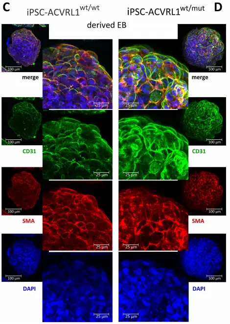
„Induction of endothelial differentiation in EBs. Depicted are confocal microscopy images of (A) ACVRL1wt/wt and (B) ACVRL1wt/mut EBs documenting the appearance of CD31+ cells after endothelial induction. Phalloidin was used to highlight the F (filamentous)-actin cytoskeleton, whilst DAPI was used to stain the nucleus. (C,D) Staining of EBs with an antibody directed against smooth muscle actin to detect possible pericyte structures.“
Reference: Xiang-Tischhauser et. al. (2023) Generation of a Syngeneic Heterozygous ACVRL1(wt/mut) Knockout iPS Cell Line for the In Vitro Study of HHT2-Associated Angiogenesis, Cells. 2023 Jun; 12(12): 1600.
Innovative 3D Tumor Model for Nanoparticle Drug Testing
Utilizing the CERO 3D Incubator & Bioreactor, tumor spheroids were generated from the human colon cancer cell line HT29-MTX-E12. This research led to the development of a microfluidic 3D intestine tumor spheroid model, enabling the testing of functionalized nanoparticles and their permeation across a mucus layer. The model supports the evaluation of nanoparticulate drug delivery systems, with a focus on improving oral drug delivery by enhancing bioavailability and targeting effectiveness
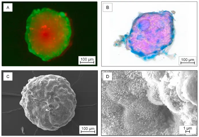
“Characterization of the 3D-tumor spheroids generated from the cell line HT29-MTX-E12. A) Live-dead-staining: Simultaneous fluorescence staining of viable (calcein-AM, green) and dead cells (propidium iodid (PI), red). B) Histological stained spheroid: Nuclear fast red – cell nucleus; Alcian blue – acid mucosubstances and acetic mucins. C) Scanning electron microscopy of a spheroid and D) and scanning electron microscopy display of the microvilli on the surface.”
Reference: Elberskirch et. al. (2021) Microfluidic 3D intestine tumor spheroid model for efficient in vitro investigation of nanoparticular formulations, Journal of Drug Delivery Science and Technology 63 (2021) 102496
Advanced Production of Myogenic Spheroids
The CERO 3D Incubator and Bioreactor generates scaffold-free myospheres. By comparing spheroids derived from primary porcine muscle cells with those from C2C12 cells, the research examines differences in morphology, growth parameters, marker expression, and myogenic potential. This comparison provides valuable insights into the production and development of myogenic spheroids for various applications.
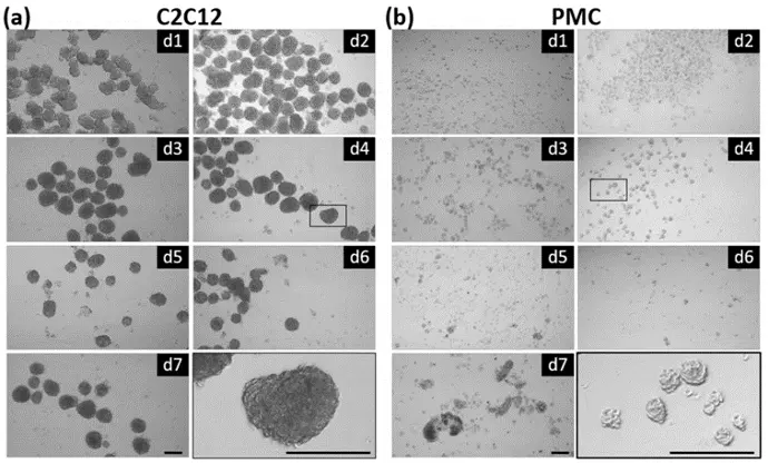
“Myosphere formation in suspension culture. C2C12 (a) and primary pig muscle cells (PMCs, (b)) were seeded (200,000 or 100,000 cells/mL, respectively) into the CERO 3D Incubator and Bioreactor (OLS) and spheroid formation was analyzed for 7 days. A considerable number of round-shaped spheroids was seen at all time points. The image on the bottom (right side) shows a magnification of spheroid(s) at Day 4. Scale bar: 200 µm“
Reference: Stange et. al. (2022) Preparation of Spheroids from Primary Pig Cells in a Mid-Scale Bioreactor Retaining Their Myogenic Potential, Cells. 2022 May; 11(9): 1453.