Application Note Cell Analysis
Detection and Quantitation of Eosinophils with Other Leukocyte Subsets in Human Peripheral Blood using the NovoCyte Flow Cytometer
Introduction
During hematopoiesis, eosinophils differentiate from myeloid cells in the bone marrow before migration into the blood. They are responsible for combating parasitic infections and play an essential role in inflammatory responses and injury occurring during an allergic response. The traditional method of identifying and quantitating eosinophils in inflammatory tissues and exudates relies on morphologic criteria and manual counting (prominent eosinophilic granules will appear as orange-red intracellular structures after H&E staining). However, there are limitations to this approach resulting from variability in sample preparation and subjective interpretation of inclusion criteria for inflammatory cells, based on morphology alone. In this application note, Dr. Paul Hutchinson and his team at National University of Singapore collaborated with ACEA Biosciences (now part of Agilent) and used a flow cytometry-based assay to identify eosinophils in human blood. This method applies a more rigorous technique for detection and allows for the exact quantitation of the cell populations of interest.
Authors
Paul Hutchinson, PhD Immunology Programme Centre for Life Sciences National University of Singapore
Brady Chung
Agilent Technologies, Inc.
Human whole blood (treated with EDTA anticoagulant) was taken from a normal donor and stained with antibodies against CD4, CD8, and CCR3. Red blood cells were then lysed in BD FACS lysing solution and the sample was washed and fixed in 2% paraformaldehyde. Human peripheral blood leukocyte samples were separately acquired using the Agilent NovoCyte flow cytometer, and a competitor's instrument. Data analysis was performed with the Agilent NovoExpress software. The wide dynamic range of detection (7.2 logarithmic decades) provided by the NovoCyte makes it easy to identify eosinophils and lymphocytes in the same plot (Figure 1A). In contrast, the limited dynamic range of the competitor’s instrument restricted the ability to identify these two cell types in the same plot. In fact, adjusting PMT voltages to accurately identify the lymphocyte population resulted in poor resolution of the eosinophils and vice versa. The NovoCyte also enabled clear distinction of monocytes from granulocytes (Figures 1A and 1B). Sample acquisition on the NovoCyte is accomplished with a syringe pump. This enables direct absolute cell counting without the addition of counting beads through selection of the appropriate statistical function in NovoExpress. Figure 1C shows the absolute count for lymphocytes, monocyte, granulocytes, and eosinophils in a representative blood sample.


Figure 1. Simultaneous analysis of eosinophils and other leukocytes in whole blood. Human peripheral blood leukocytes were used for acquisition on the Agilent NovoCyte flow cytometer and a competitor's flow cytometer. Data were analyzed using the NovoExpress software. The population highlighted in brown represents the eosinophils present in each sample (A and B). The full range scale was displayed for the data acquired on the competitor’s instrument (5-decade dynamic range). Two different leukocyte populations representing monocytes and granulocytes were also clearly distinguished for the NovoCyte data (A), but not for the competitor’s flow cytometer (B). The direct absolute count, “Abs. Count”, column in the statistical table (C) indicates the number of cells/µL for each gated subpopulation in blood.
Table 1. A summary of the antibodies used in the experiment.

The identification of eosinophils based on their FSC/SSC profile was verified using autofluorescence and expression of CCR3. Since eosinophils are intrinsically autofluorescent, the autofluorescent cells were gated in an unstained sample and backgated to an FSC/SSC dot plot (Figures 2A and 2B).
CCR3 is a chemokine receptor found primarily on eosinophils. When comparing autofluorescent cells to CD4 and CD8 lymphocytes, the autofluorescent cells had much higher levels of CCR3 than both the CD4 and CD8 positive populations (Figures 2C and 2D). Backgating these populations to an FSC/SSC plot localized the CD4 and CD8 positive cells to where lymphocytes normally reside and the autofluorescent/CCR3 positive cells to where the eosinophils were expected to reside (Figure 2E).

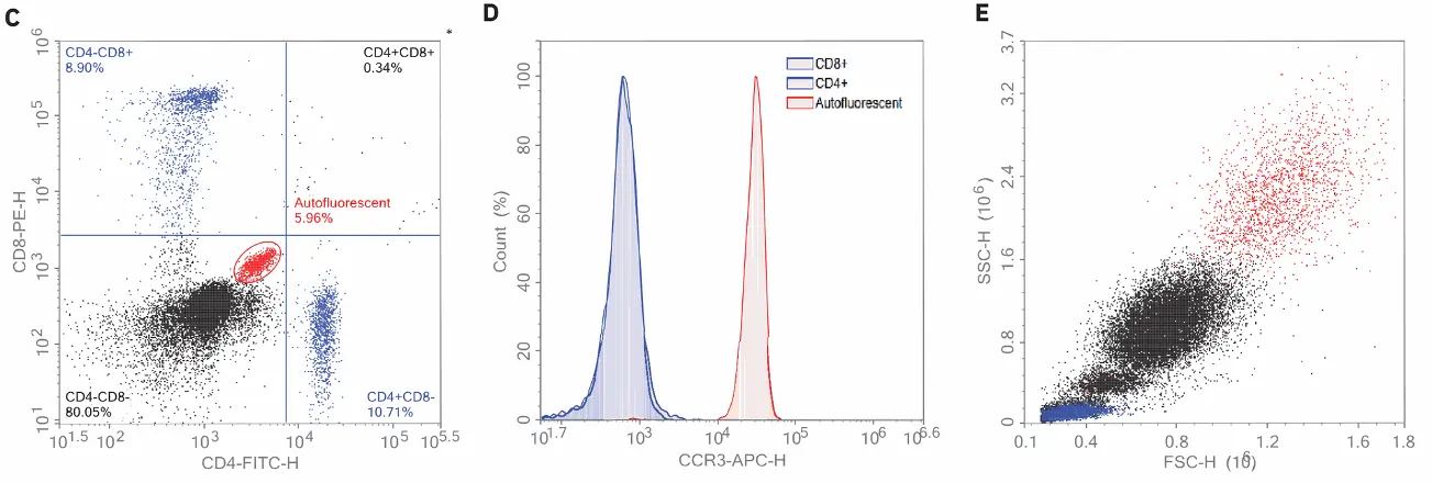
Figure 2. Identification of eosinophils using autofluorescence and CCR3. Human blood was stained with CD4, CD8, and CCR3 antibodies and acquired using the Agilent NovoCyte flow cytometer. Data were then analyzed using the NovoExpress software. Using the backgating technique, the autofluorescent cells detected using FITC/PE (A) were backgated to where eosinophils were expected in the FSC/SSC dot plot (B). Eosinophils were not only confirmed based on their strong intrinsic autofluorescence in the FITC and PE channels (C) but also through their CCR3-positive staining (D). The CD4+ and CD8+ lymphocytes are both negative for CCR3 and all cells are shown backgated to an FSC/SSC plot (E).
Conclusion
This flow cytometer-based assay offers rapid, accurate, and unbiased identification and quantitation of eosinophils in human peripheral blood, compared to conventional methods of staining and manual counting. The identification of eosinophils is based on both the FSC/SSC profile and strong intrinsic autofluorescence in the FITC and PE channels. An extra eosinophil marker, CCR3, was used to further confirm the identity of the autofluorescent population. Since eosinophils are captured together with lymphocytes, monocytes, and granulocytes, the wide dynamic range offered by the NovoCyte facilitates the resolution of all of these cell types in the same FSC/SSC plot. The separation of the monocyte and granulocyte populations were obtained based solely on their scatter profiles. Highly accurate absolute cell counts for each of these cell types was also possible, enabling the generation of additional analytic data.
References
1. Carulli, G. et al. Detection of Eosinophils in Whole Blood Samples by Flow Cytometry. Cytometry. Dec 15, 1998, 34(6), 272–9.
2. Klion, A. D; Nutman, T. B. The Role of Eosinophils in Host Defense Against Helminth Parasites. J. Allergy Clin. Immunol. 2004, 113(1), 30–7.
3. (PubMed: 14713904) Ponath, P. D. et al. Molecular Cloning and Characterization of a Human Eotaxin Receptor Expressed Selectively on Eosinophils. J. Exp. Med. 1996, 183, 2437–2448
4. Rothenberg, M. E.; Hogan S. P. The eosinophil. Annu. Rev. Immunol. 2006, 24, 147–74. (PubMed: 16551246)
5. Safdarian, N. et al. Identification of Nasal Eosinophils using Two-Photon Excited Fluorescence. Ann. Allergy Asthma Immunol. May, 2011, 106(5), 394–400. doi: 10.1016/j. anai.2010.12.019. Epub 2011 Feb 18.
6. Thurau, A. M. et al. Identification of Eosinophils by Flow Cytometry. Cytometry. Feb 1, 1996, 23(2), 150–8.
RA44139.6521527778 For Research Use Only. Not for use in diagnostic procedures. This information is subject to change without notice. © Agilent Technologies, Inc. 2020 Printed in the USA, November 24, 2020
5994-2119EN: