Biosensor Technology Meets Live Cell Imaging
Biosensor Technology Meets Live Cell Imaging

The Power of Live Cell Imaging Now Combined with the Analytical Sensitivity of xCELLigence Biosensor Technology
Exceptional versatility
Providing label-free, real-time biosensor measurements and kinetic imaging of the same live cell populations, independently or simultaneously.
Generate physiologically relevant data
Easily monitor cell health, adhesion, morphology, proliferation, and cytolysis in primary or native cells alone or in co-culture, providing unprecedented insight into cellular mechanisms and functionality.
More in-live cell imaging
The imaging platform supports three fluorescence channels, a plethora of well plate formats, an array of reporter reagents, and flexible user-defined schedules.
Very fast
Can read a 96-well plate in 15 seconds with the xCELLigence biosensor technology, and image an entire 96-well plate in 6 minutes.
Two complementary modalities, one experiment, easy workflow
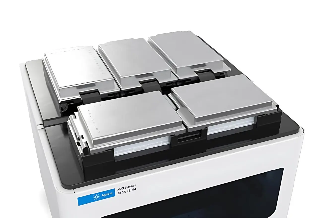
Introducing the xCELLigence RTCA eSight
The Power of Live Cell Imaging Now Combined with the Analytical Sensitivity of xCELLigence Biosensor Technology
Exceptional versatility
Providing label-free, real-time biosensor measurements and kinetic imaging of the same live cell populations, independently or simultaneously.
Generate physiologically relevant data
Easily monitor cell health, adhesion, morphology, proliferation, and cytolysis in primary or native cells alone or in co-culture, providing unprecedented insight into cellular mechanisms and functionality.
Generate physiologically relevant data
Easily monitor cell health, adhesion, morphology, proliferation, and cytolysis in primary or native cells alone or in co-culture, providing unprecedented insight into cellular mechanisms and functionality.
More in-live cell imaging
The imaging platform supports three fluorescence channels, a plethora of well plate formats, an array of reporter reagents, and flexible user-defined schedules.
Very fast
Can read a 96-well plate in 15 seconds with the xCELLigence biosensor technology, and image an entire 96-well plate in 6 minutes.
Two complementary modalities, one experiment, easy workflow
Single setup for dual measurements

Live cell imaging and real- time biosensor measurement are performed on the same cell populations to provide incisive information on cell behavior. Place plates in incubator, set up real-time data acquisition and analysis parameters, then walk away.
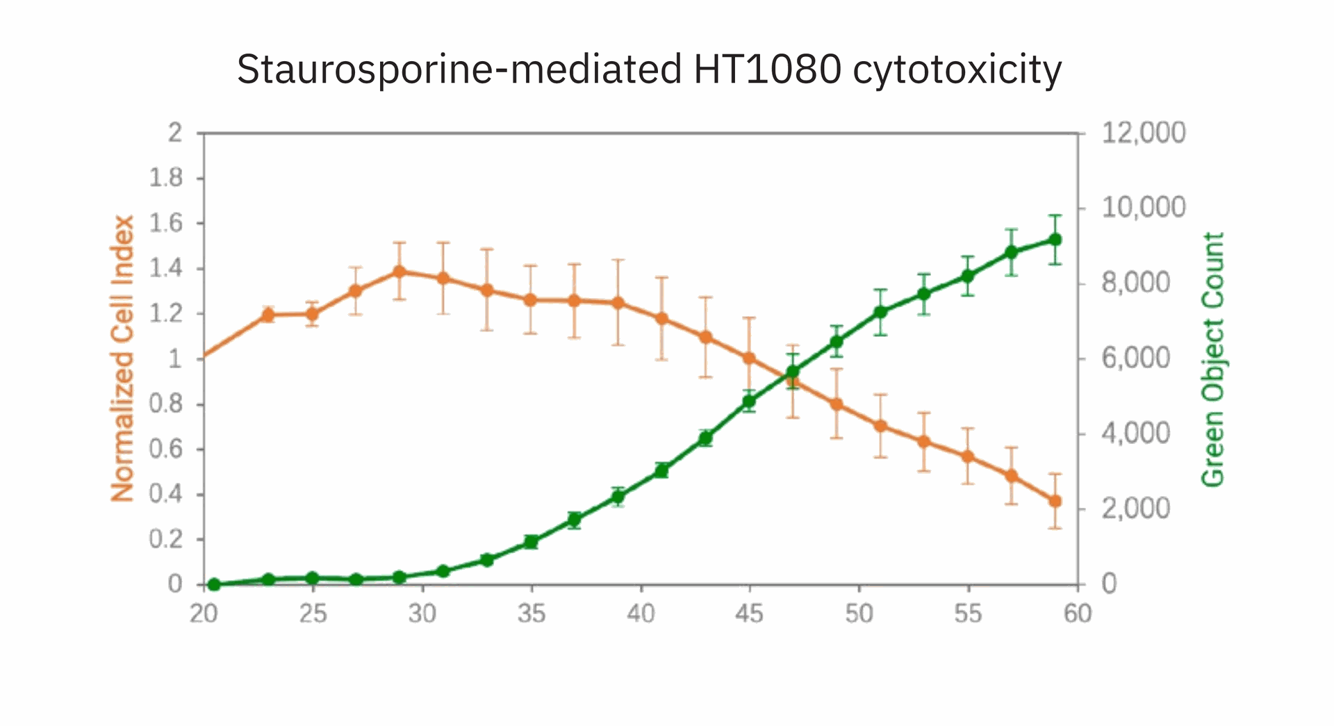
Multimodal data acquisition

Automatically acquires biosensor signal and images over time. Powerful software integrates two data types in one temporal display.
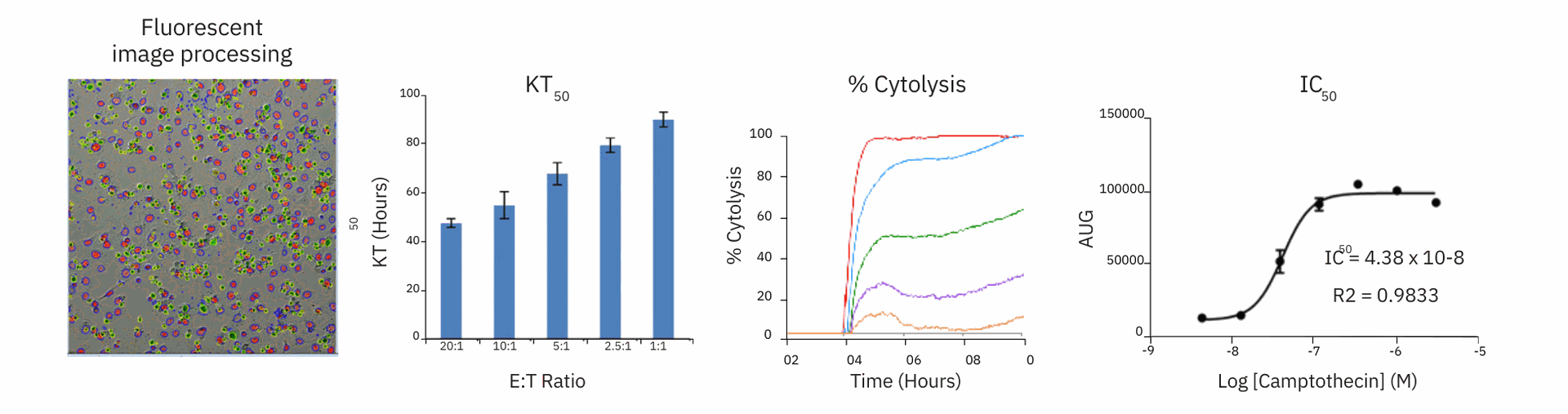
Information-rich and powerful analysis Cell analysis can be displayed and exported in diverse
formats, such as fluorescent images, KT 50 (time to reach 50% cytolysis at a given E:T ratio), % cytolysis dose response, or IC 50 dose response curves.
Functionality overview
The xCELLigence technology utilizes proprietary microtiter plates (E-Plates) embedded with gold biosensors at the bottom of each well, which serve to non-invasively quantify cell behavior. Over the course of an experiment, the biosensors monitor cell metrics such as proliferation, adhesion, morphology, migration, differentiation, and much more. The measurement is exceptionally fast and provides exquisite temporal resolution so that all relevant responses can be measured in seconds, minutes, hours, and days. In concert with the biosensor measurements, cell images can be captured in real-time, thereby providing a spatial and temporal dynamic view of the cell populations and analytically validating time-dependent cell health and behavior at an unprecedented level of details for any cell-based assay.
Broad applications
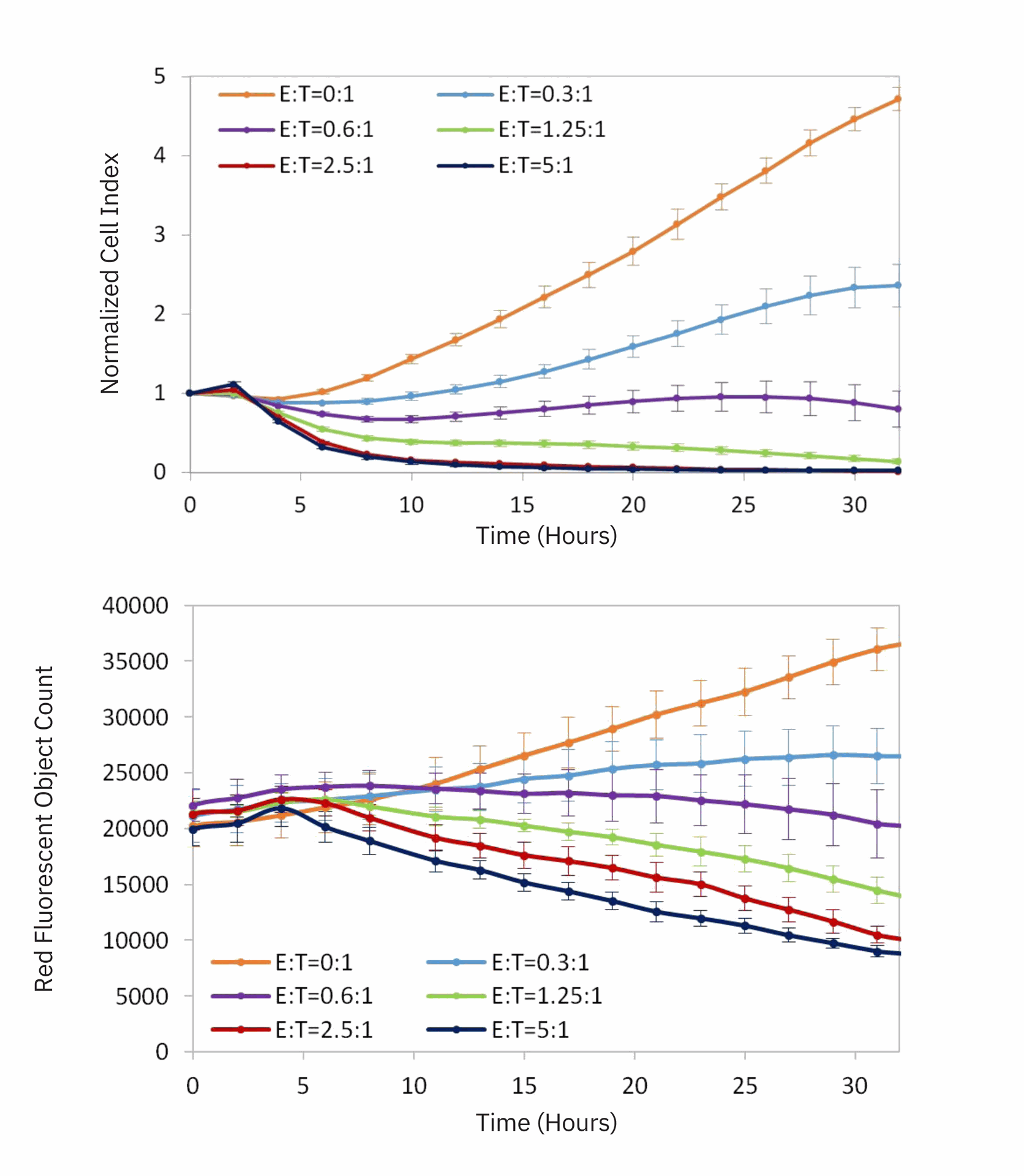
The streamlined workflow, high reproducibility, and quantitative kinetics of the eSight system makes it ideal for a wide range of cell-based assays such as proliferation, cytotoxicity, and apoptosis. The example below illustrates the monitoring of immune cell-mediated killing of cancer target cells in real-time. MCF7 breast cancer cells were transfected with a lentivirus expressing a red fluorescent protein (eLenti Red, Cat# 8711011), seeded on an E-Plate for 25 hours, and then treated with NK92 cells at different effector:target (E:T) ratios.

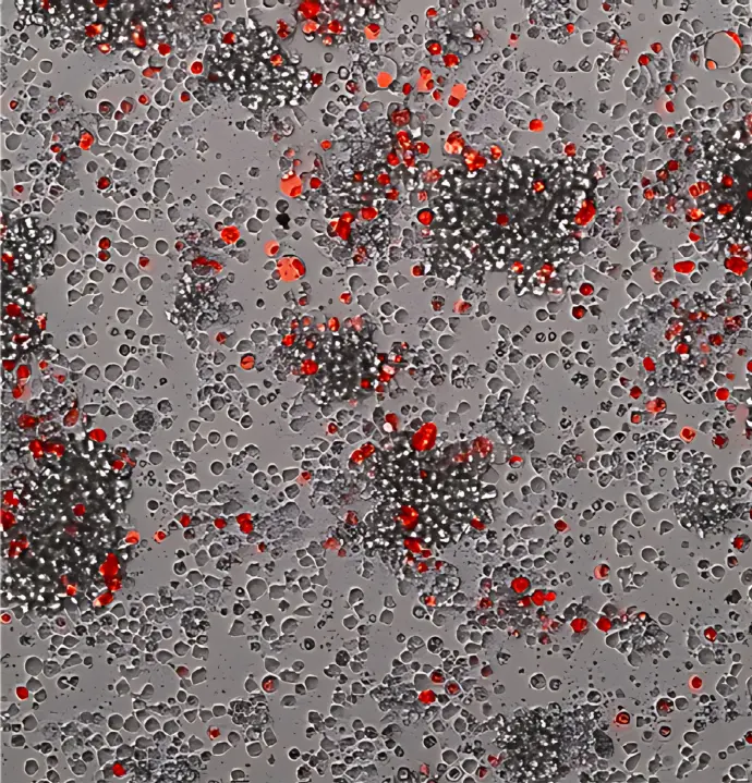
Images taken before (top left), or 12 hr (top right) and 30 hr (bottom left) after NK92 effector cells addition at an E:T ratio of 2.5:1 allow visualization of target cell (red) death over time.
For Research Use Only. Not for use in diagnostic procedures.
This information is subject to change without notice.
© Agilent Technologies, Inc. 2019
Published in the USA, August 10, 2019
5994-1219EN